

For starters, let me describe the mortality: as high as one sheep a week.
I got nervous when the owner asked me about my experience with sheep and I hastily bluffed, “A little.” I didn’t lie. I had Ruminant Medicine to back it up and clinical training in our FAC (Farm Animal Clinics Internship). But, sheep? What was I doing here?
The owner said. Well, you’re the farm veterinarian. Find out what you need to find by the end of the month.
One thing that made me really a strong practitioner was gut instincts and willingness to learn what was wrong; I pitied those helpless animals. I headed to where the sheep pen was to take a closer look. The farmhand happened to be a very amiable old man and the sheep listened only to him and obeyed his commands. He had a small stick for a cane as well as a weapon against snakes.

I didn’t have a clue. But the following week, I was excited to hear another mortality report. In veterinary medicine, we are allowed to perform preliminary necropsy as part of our training on animals belonging to or pertaining to the vertebrata (or craniata), a subphylum of chordate animals, comprising those having a brain enclosed in a skull or cranium and a segmented spinal column. This is a major taxonomic group that includes mammals, birds, reptiles, amphibians, and fish. But for every species of animal vertebrates, there was a specific procedure. I only memorized that of the dog.
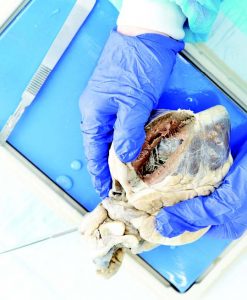
Relying on my old college necropsy manual, and with a minimum of instruments, I proceeded to perform one on a sheep in front of two other farmhands as well as curious children.
As I neared the esophagus, a huge bulge that was projecting from the left side, near the rumen, was observed and seemed to push the reticulum further forward, protruding like a distended sack of copra.
As a side note the rumen is the first of four chambers. The other chambers are named Omasum and the so-called true stomach of the ruminant or the “Abomasum,” which receives food or cud from the esophagus, third and fourth respectively, partly digests it with the aid of bacteria, and passes it to the reticulum (second chamber).
To simplify matters, the following clinical signs are observed in ruminants (sheep, cattle, goat, carabao) experiencing such rumen atony, rumen impaction as well as obstruction. At first, the animal goes off-feed (loss of appetite); on closer examination, there is loss of borborygmus (sounds from the abdomen whether gastric or intestinal) in short bowel sounds. The consequences for such uneventful signs are as follows: constipation as evidenced by absence of dung.
Eventually, these lead to obstruction ruminal acidosis then lactic acidosis in the animal’s bloodstream, if the animal is lactating as what happened to this patient. The sheep drastically reduced milk output. There also was distention of the abdomen. Oftentimes the animal is unable to rise up either due to extreme pain or production of amines such as histidine and or histamine. There is more inflammatory reaction than meets the eye and the goat’s blood vessels dilate, leading to shock. In the ensuing death of this patient, which was sudden and unnoticed as it was in a large flock. The orphaned lamb suffered malnutrition, and I desperately tried to bring in a female goat (a nanny or a doe that just gave birth or “kidded”).
This is what we call inter-species adoption for the survival of the young. This phenomena is well documented, for which there is no definitive explanation. It wasn’t easy. I tried to nurse it with infant milk preparation. Then I introduced it to the nanny goat, which for some time seemed to accommodate the lamb. However, things didn’t go well. I lost the little lamb.
Still, I was clueless for answers about these sudden deaths. That is until, I decided to cut up another recent mortality and subject it to preliminary necropsy. The vivid memories of my professors in Pathology doing this as they lectured flashed back. I remembered one of them saying, “study the dead that you will learn the intricacies of the living.”
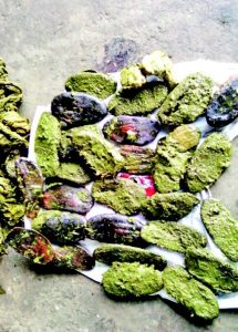
As I followed the procedure for cutting them in the prescribed manner, it yielded the answer I had long wanted to know. As I approached the rumen that was bulging, there was a motley assortment of seeds of sweet mangoes of the smaller variety which the locals in the area call “pahutan.” The number of seeds I saw per animal necropsied varied from twelve to twenty-two. Boy, I got the answers in my face.
The scenario was intriguing in that the number of seeds didn’t appeal to me as the real cause. I probed more on possible “addiction to sweets by sheep” or an article by reputable sources that had a large set of peer-reviewed materials. “Scientists have demonstrated that sugar can be an addictive substance, wielding its power over the brains of lab animals in a manner similar to many drugs of abuse. Researchers found profound behavioral changes in rats that, through experimental conditions, have been trained to become dependent on high doses of sugar. Lab animals that were denied sugar for a prolonged period after learning to binge worked harder to get it when it was reintroduced to them. They consumed more sugar than they ever had before, suggesting craving and relapse behavior.” This from Princeton University, December 11, 2008; (https://www.sciencedaily.com/releases/2008/12/081210090819.htm)
Later, in a press release, they assert that “our evidence from an animal model suggests that bingeing on sugar can act in the brain in ways very similar to drugs of abuse,” said lead researcher and Princeton psychology professor Bart Hoebel.”Princeton University released December 11, 2008, 7:13 p.m. (http://www.nydailynews.com/life-style/health/sugar-addictive-cocaine-heroin-studies-suggest-article-1.356819)
On a more sober personal note, I will try to reconcile the addiction with animal behavior in that there is a fundamental difference between rat models of animal addiction compared to a herbivore (plant-eater) like a goat. As I was tasked to give advice on how to solve the mortalities, I suggested two things. The first is to put a fence around each mango tree to disallow the sheep from bingeing on the fruits. The second was to move the pasture or grazing areas to a different place.
There is not much evidence to pinpoint addiction as a primary cause. But let me graphically demonstrate the aspect of environmental influence. When I say this, I literally mean surroundings.
Way back in 1999, a black and tan dachshund was always brought to that clinic having nervous staggers and frequent falling-over. Having observed this, I suspected the poor dog was intoxicated with something alcoholic in nature. I immediately did a physical examination.
I smelled the unmistakable scent of alcohol in its breath. I directed my history-taking to the human companion. As I probed for more information, I learned that the dog was allowed too frequently in the company of its human owner, who was in the habit of drinking beer. I was skeptical and asked, “Is it possible that your boss allows or even encourages this dog, still a puppy at the time of about four to six months old, to drink beer?” The reply was in the affirmative.
From then on I warned the house help to inform the owner of the dangers as well as consequences of chronic intake as well as inebriation to alcohol by a dog. That unfortunately did not deter the owner from giving his dog a drink once again. I had to admonish the owner with a chilling letter telling him of the applicable provisions of R.A. 8485.
This appeared in Animal Scene magazine’s February 2018 issue.


