

Leptospirosis is a worldwide infectious disease. Known as ‘lepto’, it is caused by bacteria from the genus Leptospira.
The most prevalent serovars are believed to be Grippotyphosa, Pomona, and Bratislava. Within each species, Leptospira serovars are recognized distinctly, with about 250 different serovars of pathogenic Leptospira that are identified based on surface antigens.
A serovar is a distinct variation within a species of bacteria or virus among immune cells of different individuals. (Wikipedia)
Throughout the world, it affects all warm-blooded mammals physically and is manifested in a wide variety of clinical effects — mild, subclinical, and multiple organ failure that may eventually lead to death, which is still quite common in unvaccinated dogs in the Philippine setting.
The clinical experience with this disease is almost similar among different clinics. In almost all cases, survival is at 70 to 85 percent, given that treatment is provided before clinical signs as jaundice, fever, abdominal pain, and vomiting are seen.
Cats are considered historically resistant but have been experimentally infected and do produce antibodies against Leptospira when vaccinated.
Limitations of clinical diagnosis without confirmatory diagnosis
In the early days of veterinary practice and at the time I started Companion Animal Medicine, diagnosing Leptospirosis was purely clinical. Whenever a veterinarian attended to a gravely sick dog who had jaundice and loss of appetite, it would send shivers down my spine.

The affected animal wouldn’t move and would seem depressed. If the owner mistook it as a transient loss of appetite, it would be disastrous, to say the least. The animal would then have fever and would eventually vomit blood.
I also observed that their breath would smell like urine (which we would call uremic fetor). This would mean the kidneys weren’t functioning well. Jaundice, on the other hand, indicates the liver is manifesting overt disease.
Present-day diagnostics
Nowadays, many veterinarians use test kits like enzyme-linked immunosorbent assay (ELISA), microscopic agglutination tests (MAT), culture and isolation, dark-field microscopy, fluorescent antibody techniques, and a host of many other non-specific tests, such as serum chemistry, urinalysis, and antibody titer tests.
Common examination procedures include abdominal palpation and routine measurements of temperature, pulse, and respiratory rate. It is not a good sign if there is a yellow discoloration of the mucous membranes of the gums, nostrils, and genitals.
Fight against lepto
There are available bivalent and quadrivalent canine vaccines against leptospirosis caused by icterohaemorrhagiae, canicola, pomona, and grippotyphosa. Consult your veterinarian for the diagnosis and medical management regarding this fatal and contagious disease.
Leptospirosis is associated with reversible kidney injury and poses an 80 percent chance of survival. However, the longer one had to wait before bringing an animal to the clinic, the worse the prognosis.
The acute phase requires intravenous use of penicillins, together with supportive fluid treatment and correction of possible existing electrolyte and acid-base imbalances. Supportive treatment includes the use of drugs that prevent vomiting, gastrointestinal protectants, phosphate binders that help prevent the worsening of kidney function, liver protectants, and herbal meds.
Prompt treatment with intravenous medication to eliminate the pathogen even before confirmatory laboratory diagnosis is a must.
Save the kidneys!
The prevention of acute kidney injury is crucial in instituting a reversal. Early diagnosis and treatment are key to a higher probability of survival.
We also want to terminate the shedding of leptospires. Regardless of the serovar involved, treatment protocol is the same. Knowledge of the infecting serovar is essential in epidemiological studies as well as effective development of vaccines.
There is currently no tailor-fit treatment intended to address specific serovars.
General precautions
1. Observe barrier precautions
2. Avoid exposure to excreta, particularly urine or blood
3. Dogs must have designated urination areas where cleaning and disinfection
are easy
4. Owners should wear latex gloves when cleaning up urine
5. Wash hands after handling a dog until the course of antibiotic therapy is
complete
Extra musings
This jaundiced Alaskan Malamute named Aswang was recommended for ethical euthanasia. The previous attending vet had no other solutions to cure him. The dog is still alive today.
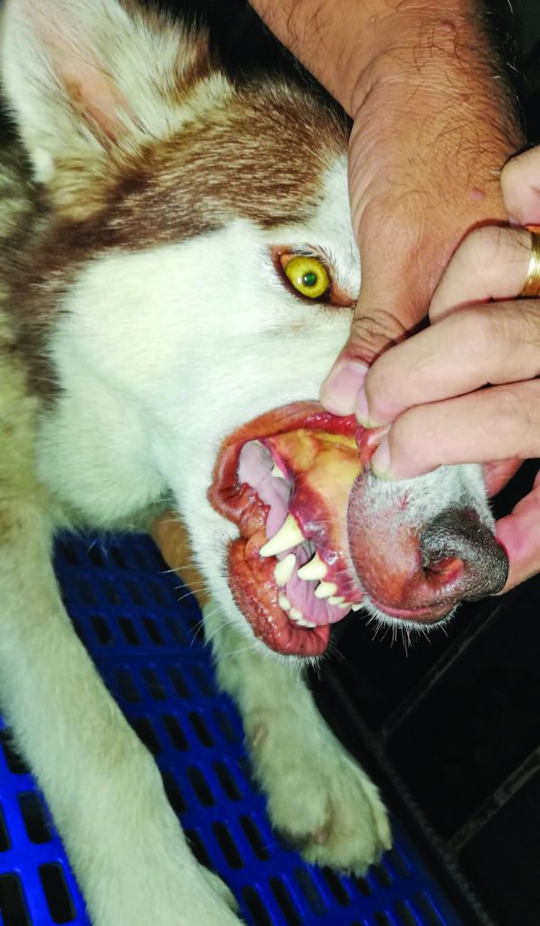
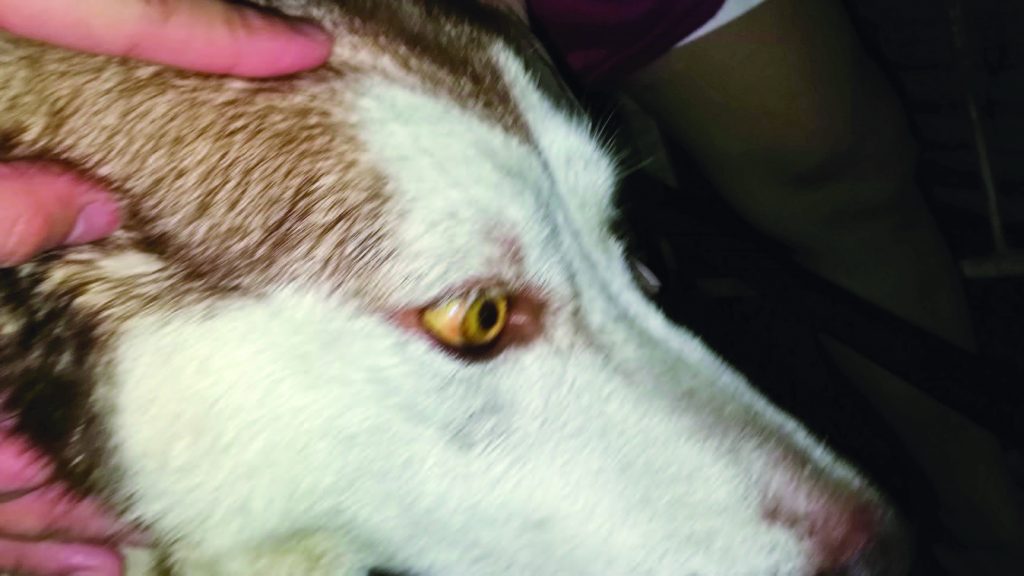
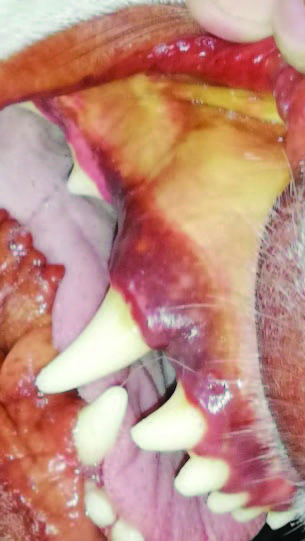
A clinician nowadays would not be too confused about the diagnosis, given the odor of his breath, as well as the jaundiced eyes and gums. There was a slight decrease in platelet count and an unconvincing immunochromatographic test for Ehrlichiosis.
I had to save the dog fast. The dog recovered after two weeks of treatment. I felt relieved that that the prompt treatment of leptospirosis was preferred over lab results, which would have come too late. The dog is still undergoing treatment for possible subclinical infection.
Dr. Emmanuel Macapagal is the 2000-2001 president of the Philippine Animal Hospital Association, Inc.
This appeared in Animal Scene magazine’s May 2019 issue.
Related stories:
– How often should your pet see the vet?
– Vetiquette: Proper behavior at the pet clinic
– Guidelines for pet owners, vets, civic groups


